3D imaging reveals secret fibers that connect tendons to bone

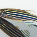
The Achilles tendon is a powerful body part connecting calf muscle to heel bone, enabling springing leaps and hurdles that make sports stars seem superhuman. But exactly how it connects to the bone hasn’t been fully understood — until now. Researchers from multiple disciplines took a microscopic look and found an extremely thin tissue layer that enables the tendon’s great expansive strength.
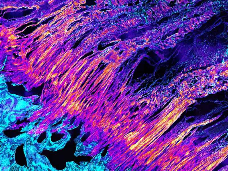
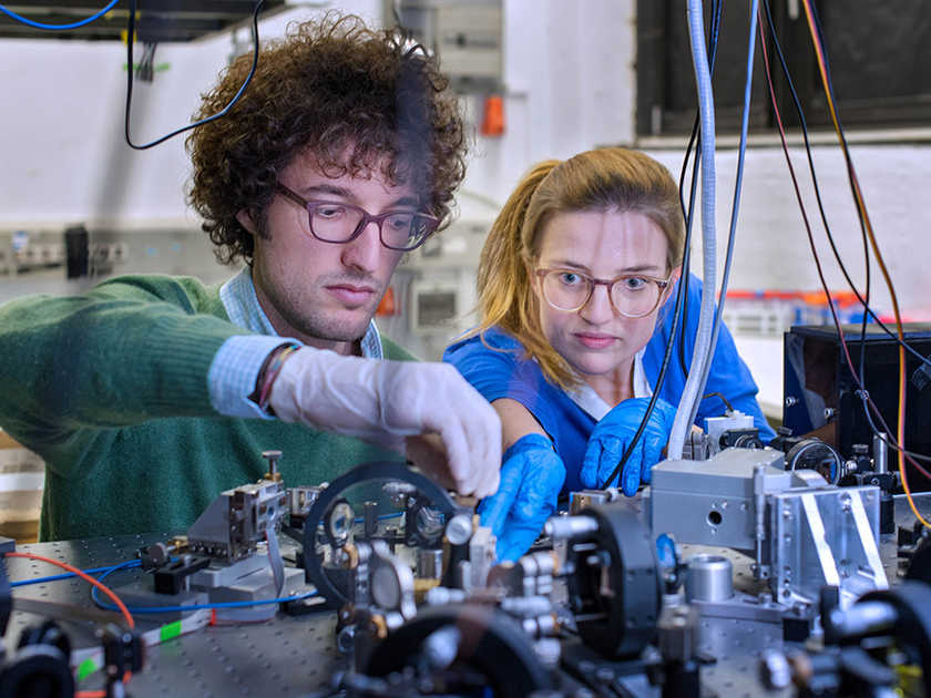
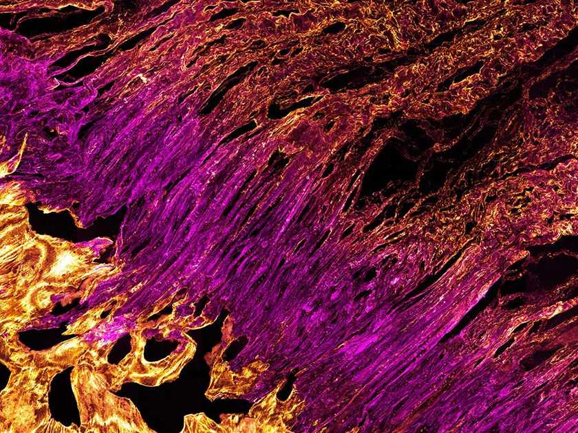
A team of biochemists and biophysicists from the Technical University of Munich headed by physician Dr. Rainer Burgkart applied a sequence of investigatory methods to get a close look. First, a series of multiscale microscopic photos of the boundary layer between tendon and bone were combined into a single large image, which revealed the former’s tissue splitting into dozens of tiny fibers. Fluorescent antibodies lit up some specific proteins in the layer, meaning its biochemical makeup is distinct from the tendon’s. Finally, applying weight to the tendon showed different fibers acting to stabilize it depending on the direction it was moved.
Sure, there have been innovative stabs at replacing tendons with nanotech, but the the fancy new synthetic parts will still have to be securely fastened to your remaining organic ones. Understanding precisely how this tendon connects to bone has several applications for future medicine, like attaching existing tendons to orthopedic implants. It’s also promising for materials research, which could lead to new methods for connecting soft and hard synthetic parts.
(25)